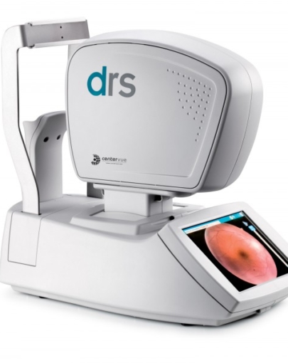
Description
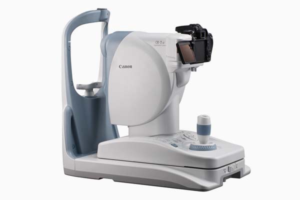
The Canon CR-2 Digital Non-Mydriatic Retinal Camera incorporates the latest in Canon retinal imaging technology and enhancements in a compact and light design. It can easily be installed and takes up minimal office space and, for added convenience, can remain mobile for easy transportation when needed. The illuminated control panel on the CR-2 allows medical staff to easily navigate operations in darkened rooms. The CR-2 can help reduce energy costs in medical facilities with its energy-efficient design contributing to a lower total-cost-of-ownership.
Features
-
Compact and Lightweight
The small design of the CR-2 provides the smallest footprint of any Canon retinal camera to date and is portable when needed. Canon instrument tables (sold separately) may comfortably fit a CR-2 and a Canon Auto-Tonometer (sold separately). Keeping the Canon machines next to one another helps expedite the exam process. -
Mobile Retinal Imaging Solution
The mobile retinal imaging solution is like having onsite Teleretinal Screenings in a box! Designed exclusively for the CR-2 Non-Mydriatic Retinal Camera, the custom, hardshell travel case is equipped with top and side carry handles and wheels making the CR-2 Retinal Camera easy to carry to and from locations where stationary retinal imaging equipment is unavailable. The custom travel case accommodates (each sold separately) the CR-2 Non-Mydriatic Retinal Camera with dedicated camera for CR-2, laptop (max. dimensions (HxWxD) 1.47 x 16.4 x 10.7 inches), external fixation lamp, mouse, power cord and USB cable. -
Canon EOS Camera Technology
High quality diagnostic images are obtained using a dedicated EOS camera for retinal imaging which incorporates a large, high-definition CMOS sensor. -
High Resolution Images
The CR-2 provides high resolution images, making it easier for the diagnosis and detection of a wide range of ocular conditions and diseases. -
Digital Filter Processing
Red-Free and Cobalt digital filters are included and provide enhanced screening exams. Red-Free is used for evaluating the retinal nerve fiber layer (RNFL) and vascular structure of the retina associated with documenting Glaucoma, Diabetic Retinopathy or Hypertension. The Cobalt filter is also used for evaluating the RNFL, as well as, Optic Disc and Optic Disc Drusen. Additionally, Green (Vascular view) and Red channel (Choroid view) digital filter views are also included. -
White LED Flash
Using white LED flash light as a light source saves on power consumption and energy costs. -
Low Flash Photography
The low flash intensity of the CR-2 minimizes miosis, thus shortening the time required for taking multiple view exams or stereo images. The reduced brightness improves patient comfort and reduces the “ghost” image the patient sees after an exposure. -
Automatic Exposure Function
The observation light automatically optimizes the light brightness and flash intensity. -
Ergonomic Control Panel
The simplified design of the control panel can be easily handled by a trained examiner. A one-hand joystick positions the camera for a desirable point of view. In darkly lit rooms, the operation panel illuminates for easier navigation. The 35 mm working distance between the patient and the operator allows easy access to the patient’s eyes should they need assistance with opening their eyes. -
Retinal Imaging Control Software
Using the Canon Retinal Imaging Control Software (RICS), images can be stored, viewed, processed and sent to a printer. The Canon RICS complies with the DICOM®* Standard. Images may be stored as DICOM or JPEG files and sent to a printer. For more information, visit Retinal Imaging Control Software.*DICOM is a registered trademark of the National Electrical Manufacturer Association for its standard publications relating to digital communications of medical information.

