


Description
The ZEISS Stratus OCT Model 3000 (Stratus OCT) enables examination of the posterior pole of the eye at an extremely fine spatial scale, without surgical biopsy or even any contact with the eye.
The name Stratus OCT (derived from “stratum,” Latin for “layer”) refers to its unique ability of direct cross-sectional imaging of the layers of the retina. The Stratus OCT minimizes patient discomfort as it permits detailed examination of the retina and optic nerve head at the office or clinic. The Stratus OCT facilitates diagnosis and management of retinal diseases and glaucoma.
Intended Use
The Stratus OCT is intended for use as a diagnostic device to aid in the management of ocular diseases.
Indications for Use
The Stratus OCT is a non-contact, high resolution tomographic and biomicroscopic imaging device. It is indicated for in vivo viewing and axial cross-sectional imaging and measurement of posterior ocular structures, including retina, retinal nerve fiber layer, macula, and optic disc. It is intended for use as a diagnostic device to aid in the detection and management of ocular diseases including, but not limited to, macular holes, cystoid macular edema, diabetic retinopathy, age-related macular degeneration and glaucoma.
Stratus OCT System Described
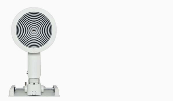
The Stratus OCT is a computer-assisted precision optical instrument that generates cross sectional images (tomograms) of the retina with ≤ 10 micrometers axial resolution. It works by using an optical measurement technique known as low-coherence interferometry.
How The Stratus OCT Works
The principle of operation of interferometry is analogous to ultrasound, except that it uses light rather than sound. The difference permits measurement of structures and distances on the ≤ 10 micrometer scale, versus the 100-micrometer scale for ultrasound. Another important difference is that optical interferometry does not require contact with the tissue examined, unlike ultrasound.
The Stratus OCT contains an interferometer that resolves retinal structures by measuring the echo delay time of light that is reflected and backscattered from different microstructural features in the retina. The Stratus OCT projects a broad bandwidth near-infrared light beam (820 nm) onto the retina from a super luminescent diode. It then compares the echo time delays of light reflected from the retina with the echo time delays of the same light beam reflected from a reference mirror at known distances. When the Stratus OCT interferometer combines the reflected light pulses from the retina and reference mirror, a phenomenon known as interference occurs. A photodetector detects and measures interference. Although the light reflected from the retina consists of multiple echoes, the distance traveled by various echoes is determined by varying the distance to the reference mirror. This produces a range of time delays of the reference light for comparison.
The Stratus OCT interferometer electronically detects, collects, processes and stores the echo delay patterns from the retina. With each scan pass, the Stratus OCT captures from 128 to 768 longitudinal (axial) range samples, i.e., A-scans. Each A-scan consists of 1024 data points over 2 mm of depth. Thus the Stratus OCT integrates from 131,072 to 786,432 data points to construct a cross sectional image (tomogram) of retinal anatomy. It displays the tomograms in real time using a false color scale that represents the degree of light backscattering from tissues at different depths in the retina. The system stores the scans you select for later analysis.
How It Works for You
The Stratus OCT delineates intraretinal, cross-sectional anatomy with axial resolution of ≤10 micrometers and transverse resolution of 20 micrometers. The Stratus OCT software package includes 19 scan acquisition protocols and 19 analysis protocols. Together they enable you to analyze the optic disc, retinal nerve fiber layer and the macula with a single instrument. The Stratus OCT facilitates the evaluation of both glaucoma and vitreoretinal disease. The Stratus OCT video camera enables you to view the patient’s fundus as you work, and to store video and scan images together. The data management system enables you to keep Stratus OCT histories, so you can monitor patients over time. You can archive images and data on rewritable DVD-RAM disks. The inkjet printer enables you to generate color hard copy.
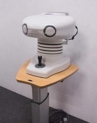
Stratus OCT System Hardware
The Stratus OCT system hardware consists of the Patient Module; the Computer Unit; the Flat Screen Video Monitor; the keyboard, mouse and color inkjet printer. The hardware mounts on a wheelchair accessible motorized power table. The table accommodates elevation adjustment to each patient’s height. The illustration below labels hardware elements.
Stratus OCT Detials
The Stratus OCT incorporates optical coherence tomography technology to provide comprehensive imaging and measurement of glaucoma and retinal disease. Stratus OCT is the gold standard in vivo imaging device for the posterior segment and offers proven reproducibility for disease management.
The Stratus OCT provides real-time cross-sectional images and quantitative analysis of retinal features to optimize the diagnosis and monitoring of retinal disease and for enhanced pre- and post-therapy assessment. The device is beneficial for evaluation of cataract patients, pre- and post-operatively, and for the assessment of early signs of glaucoma and glaucomatous change.
Retinal Nerve Fiber Layer (RNFL) Analysis
Circular scans around the optic nerve head capture RNFL measurement of the peripapillary region. Analyses provide comparison of measurements to a normative database, demonstration of asymmetry and serial analysis.
Optic Nerve Head Analysis
Radial line scans through the optic disc create cross-sectional and topographical data. The key analysis parameters include cup-to-disc ratios and horizontal integrated rim volume.
Macular Thickness Analysis
Radial line scans provide cross-sectional images and analyses revealing retinal layers and macular condition. The change analysis feature illustrates change over time.
Specifications
- Tomographic Imaging
- Purpose: Cross sectional imaging of fundus
- Signal Type: Optical scattering from tissue
- Signal source: Super Luminescent Diode (SLD), 820 nm
- Optical Power: ≤ 750 microwatts at cornea. SLD current will shut off if a safety circuit is activated (upon scanner failure)
- Spot Size at Retina: 20 μm in tissue
- Longitudinal/Axial Resolution: ≤ 10 μm in tissue
- Transverse Resolution: 20 μm in tissue
- Scanners: Galvanometric mirrors
- Scan Patterns: Repeat, Line, Circle, Raster Lines, Cross Hair, Radial Lines, Macular Thickness Map, Optic Disc, Proportional Circle, Concentric 3 Rings, RNFL Thickness (3.4), Nerve Head Circle, RNFL Thickness (2.27xdisc), X-Line, RNFL Map, Fast Macular Thickness Map, Fast Optic Disc, Fast RNFL Thickness (3.4), Fast RNFL Map
- Scan Pixels: Adjustable from 131,072 to 786,432 (1024 longitudinal x 128 to 768 transverse)
- Scan Speed: 400 A-scans per second
- Scan Acquisition Time: Approx. 0.32 seconds (128 A-scans minimum.) to approx. 1.92 seconds (768 A-scans maximum.)
- Longitudinal (Depth) Scan Range: 2 mm in tissue