

Description
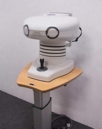
Backed by 30 years of industry-leading experience, the CR-1 Digital Non-Mydriatic Retinal Camera delivers ultra-high resolution images in an ergonomic, easy to use design while also providing quicker, more comfortable exams for the patient. The CR-1 features true 45-degree imaging, 2X magnification, intuitive controls, low flash mode, illuminated operation panel, one-handed joystick operation, efficient workflow and more.
Benefits of Low Flash Intensity
The efficiency of non-mydriatic retinal exams heavily depends on a patient’s darkness adaptation period. The low flash intensity of CR-1 Mark II minimizes miosis, thus shortening the time required for taking multiple pictures such as binocular and stereo images. The reduced brightness induces more patient compliance, leading to speedy and accurate exams for high throughput screening.
High-Quality Retinal Imaging
Canon’s expertise in the field of imaging technology supports CR-1 Mark II’s exceptional digital retinal imaging capabilities. High-resolution diagnostic images of the retina are extremely detailed and clear, a necessity to effectively detect and monitor ocular conditions such as diabetic retinopathy, glaucoma, and macular degeneration.
45-degree Angle of View
The CR-1 Mark II features an optical system that achieves high-resolution diagnostic images at a 45° angle of view suitable for health check-ups and screening.
2X Digital Magnification
“2X” mode provides a magnified view of the retina for viewing the details of the region of interest. Sharp images are made possible by the attached EOS digital SLR camera’s high pixel count.
Ergonomic Controls
CR-1 Mark II has an ergonomic design to simplify operation for the user. A one-hand joystick repositions the camera to the exact view desired. The illuminated operation panel, featuring easy-to-understand control options, has been designed for quick adjustment and selection of the necessary settings. A short reaching distance between the patient and the operator allows closer personal interaction and easy access to the patient’s eyes.
Safe, Secure Patient Positioning
An adjustable motorized chin rest, forehead guard, and front protection cover perfectly situate the patient securely in position for the exam while ensuring their safety during operation. An optional external fixation target is also available.
Easy Alignment and Focusing
Two simple steps are all it takes to capture a clear image. Step 1 is the aligning of two halves of a split pupil image, followed in Step 2 by the adjustment of split lines and working distance dots in the retina display. This ensures that the correct focus and working distance are achieved for the sharpest images possible.
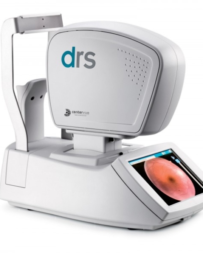
Retinal Imaging Control Software
The bundled Retinal Imaging Control Software for the CR-1 Mark II puts tools for comprehensive study management, image capture controls, and easy viewing at your fingertips. The intuitive graphical interface is simple and straightforward to use. The PC-based software provides quick input and access to all information and images required to assist in your diagnosis, and data can be easily saved to various external media. For more information, visit the Retinal Imaging Control Software webpage.
Printers and Projectors
If your imaging requirements include color or black-and-white prints in a variety of sizes, Canon printers offer the terrific blend of print quality, ease-of-use, speed, and cost-effective operation. For teaching facilities and conference rooms, Canon’s high-quality projectors provide LCOS (liquid crystal on silicon) technology, which is engineered to provide exceptional color, intricate details, and easy-to-read type.